Prof Wong Pak Kin in the Faculty of Science and Technology of the University of Macau (UM) and his doctoral student Yan Tao in the Department of Electromechanical Engineering have developed an intelligent system that can successfully distinguish pneumonia caused by the novel coronavirus (COVID-19) from other common types of pneumonia, at a speed that is nearly 60 faster than radiologists, bringing a new feasible solution for pneumonia detection. The study has been published by the international science journal Chaos, Solitons & Fractals.
COVID-19 infection is typically confirmed through reverse-transcription polymerase chain reaction (RT-PCR). However, RT-PCR tests have many problems, such as insufficient supply of the RT-PCR kit, the amount of time required, and a high rate of false negatives. These can impede prompt diagnosis and increase the chances of the spread of the virus. Many experts have proposed using chest computed tomography (CT) to diagnose suspected cases of COVID-19 infection, because CT can detect infection even during the first stage. CT diagnosis has a very high degree of accuracy and can provide more clinical information, but CT images require manual identification of radiographic features. The rapid growth in the number of COVID-19 patients and the necessity for each patient to undergo multiple CT scans (more than 100 slices per scan) have led to a large number of CT images, presenting an enormous challenge for radiologists, especially in areas hit hardest by the pandemic.
In order to address this issue, Prof Wong and Yan Tao worked with researchers at other institutions in Hubei province, namely Wang Jiangtao, deputy director of the Department of Radiology at Xiangyang Central Hospital and Doctor Ren Hao at the same hospital; Li Yang, deputy director of the Department of Radiology at Xiangyang First People’s Hospital; and Wang Huaqiao, a surgeon in the Division of Surgery at the same hospital. They retrospectively collected data on 206 patients with confirmed positive RT-PCR for COVID-19 and their 416 CT scans, as well as data on 412 patients with non-COVID-19 pneumonia and their 412 chest CT scans with clear signs of pneumonia. Based on these CT images, they developed an automatic diagnosis system based on a multi-scale convolutional neural network. The verification results have shown that after being fed a limited amount of data, the intelligent diagnosis system can successfully distinguish COVID-19-caused pneumonia from other common types of pneumonia. Its diagnostic ability is comparable to that of experienced radiologists, but the diagnosis speed is nearly 60 faster. This brings a new feasible solution for pneumonia detection. Prof Wong’s team hopes to expand the functions of the system. They are working to develop a multi-type pneumonia diagnosis algorithm and a new coronavirus pneumonia severity prediction algorithm, which is expected to be completed soon. The intelligent system will be able to distinguish between normal lungs and lungs infected with five common types of pneumonia, thereby predicting the severity of the condition of COVID-19 patients.
The related paper, titled ‘Automatic Distinction between COVID-19 and Common Pneumonia using Multi-Scale Convolutional Neural Network on Chest CT Scans’, has been published by the international science journal Chaos, Solitons & Fractals, a SCI Q-1 journal, only one month after its submission.
澳門大學科技學院王百鍵教授及機電工程系博士生晏濤開發一種基於多尺度卷積神經網絡的智能自動診斷系統,能成功區分新型冠狀病毒肺炎和其他常見的肺炎,診斷速度比醫生快將近60倍,為肺炎檢測帶來新的可行方案。該研究剛獲國際科學期刊Chaos, Solitons & Fractals發表。
新冠病毒肺炎一般通過深喉唾液核酸測試來確認,但核酸測試也存在不少缺點,例如供應不足,費時且假陰性率高等問題,可能導致患者無法及時診斷,更可能導致病毒擴散。目前,眾多專家已建議使用胸部電腦斷層掃描(CT)來診斷可疑病例,因為即使在發病初期,也可以透過胸部電腦斷層掃描來檢測。CT診斷準確性高且可以提供與治理有關的詳盡資訊,但胸部電腦斷層掃描圖像則需要人手識別其特徵,加上患者眾多及每位患者的多次CT掃描皆產生了大量的CT圖像(每次掃描平均產生過百片圖像),這對身處疫情嚴峻地區的放射科醫生來說是一個重大挑戰。
為此,王百鍵教授及晏濤於疫情初期便與湖北省襄陽市中心醫院放射科的副主任王江濤、醫師任浩、襄陽市第一人民醫院的放射科副主任李陽以及普外科主治醫師王華僑合作研究,取得了這兩間醫院的206個核酸檢測為陽性的個案及他們416組胸部電腦斷層掃描圖像。另一方面,他們在醫院內也取得了412組沒有新型冠狀病毒但只有普通肺炎的胸部電腦斷層掃描圖像。
基於這些少量CT圖像,研究團隊研發一種基於多尺度卷積神經網絡的自動診斷系統。驗證結果表明,在有限數量的訓練數據下,該智能診斷系統能成功區分新型冠狀病毒肺炎和其他常見的肺炎,其診斷能力與經驗豐富的放射科醫生相當,但診斷速度卻比醫生快將近60倍,這為肺炎檢測帶來新的可行方案。此外團隊還進一步拓展該系統的功能,他們開發的多類肺炎診斷算法及新型冠狀病毒肺炎嚴重性預測算法已接近完成。不久後,該智能系統將可具有區分正常肺部與五種常見肺炎及對新冠病毒肺炎患者進行嚴重性預測的能力。
研究論文《運用多尺度卷積神經網絡從胸部電腦斷層掃描圖像中自動區分新型冠狀病毒肺炎和普通肺炎》(Automatic Distinction between COVID-19 and Common Pneumonia using Multi-Scale Convolutional Neural Network on Chest CT Scans)剛獲國際科學期刊Chaos, Solitons & Fractals發表,此論文從遞交至正式發表僅用了一個月。Chaos, Solitons & Fractals 是SCI一區期刋。
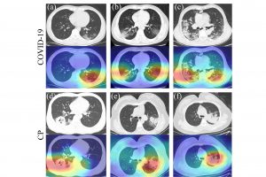
CT scan images of patients with COVID-19-caused
pneumonia and patients with regular pneumonia
together with region of lesions generated by the
intelligent diagnosis system
新冠肺炎病者及普通肺炎病者的胸部電腦斷層掃描圖像
及智能診斷系統產生的肺炎部位識別圖
Prof. Wong Pak Kin of FST
科技學院王百鍵教授
Ph.D. student from the Department of
Electromechanical Engineering, Yan Tao
機電工程系博士生晏濤


